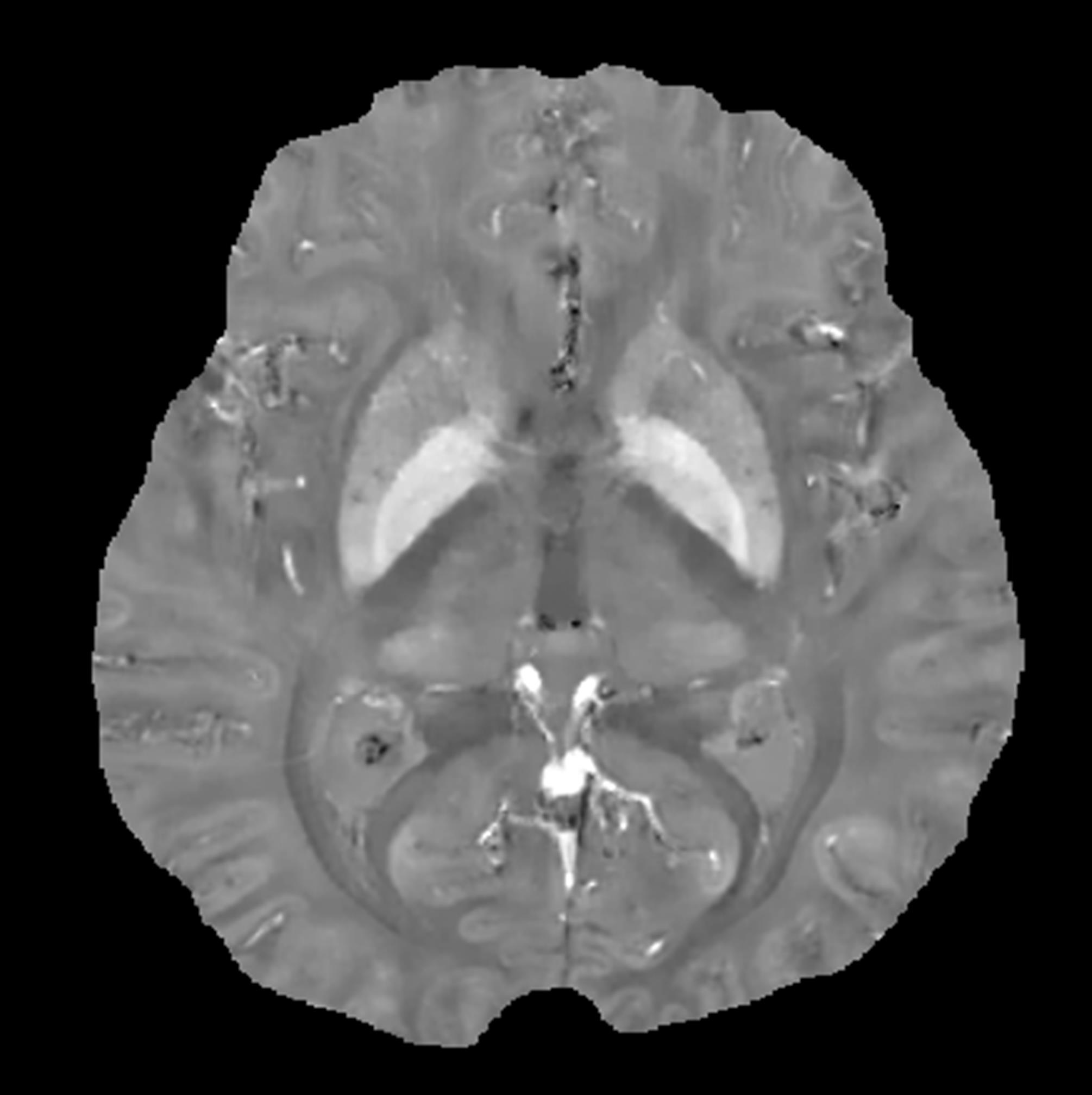
 Recently our group has pioneered quantitative susceptibility mapping (QSM), a new
fundamental approach to MRI. QSM solves the inverse problem from the magnetic field
to local magnetization source for studying tissue magnetic susceptibility. Prior
to QSM, MRI practice typically utilizes magnitude images and mostly discards phase
images. MRI phase image allows estimation of the local magnetic field, which is
a convolution of the magnetic dipole kernel with the tissue magnetic source. The
deconvolution or inversion from the magnetic field to magnetic source (tissue magnetic
susceptibility in MRI) is needed to study tissue, but this inverse problem is ill
posed due to zeroes in the dipole kernel. We have developed an accurate and robust
Bayesian approach that solves this inverse problem for the first time, therefore
establishing QSM technology. QSM generates a new contrast that is a valuable addition
to the traditional contrasts in MRI. QSM enables quantitative mapping of exogenous
contrast agents in molecular MRI (R01CA178007)
and contrast enhanced MRI, as well as quantitative mapping of endogenous contrast
agents, including iron and calcium depositions and deoxyhemoglobin, that are fundamental
for investigating organ functions and disease pathophysiology. We are currently
working with neurological clinicians and neuroscientists to develop clinical applications
of QSM (R01EB013443). Potential clinical
applications include mapping iron deposition in neurodegenerative diseases, including
Parkinson and Alzheimer diseases and multiple sclerosis (R01NS090464)
, assessing blood products
in cerebral microhemorrhage and hemorrhagic stroke
(R01NS072370), and evaluating oxygen metabolism in ischemic strokes and
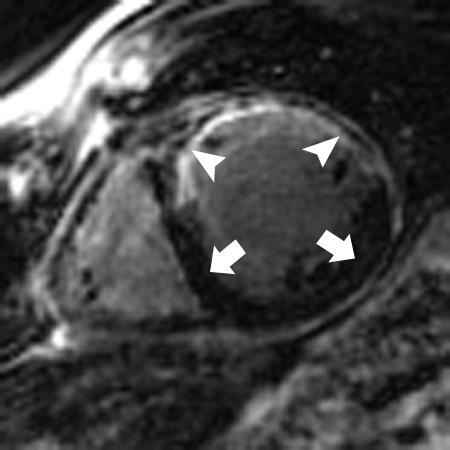
tumors. We are also investigating other important applications of QSM, including
mapping bone mineralization and iron deposition in the liver and heart, and extending
QSM to a tensor version to study myelin's unique diamagnetic susceptibility anisotropy.
Recently our group has pioneered quantitative susceptibility mapping (QSM), a new
fundamental approach to MRI. QSM solves the inverse problem from the magnetic field
to local magnetization source for studying tissue magnetic susceptibility. Prior
to QSM, MRI practice typically utilizes magnitude images and mostly discards phase
images. MRI phase image allows estimation of the local magnetic field, which is
a convolution of the magnetic dipole kernel with the tissue magnetic source. The
deconvolution or inversion from the magnetic field to magnetic source (tissue magnetic
susceptibility in MRI) is needed to study tissue, but this inverse problem is ill
posed due to zeroes in the dipole kernel. We have developed an accurate and robust
Bayesian approach that solves this inverse problem for the first time, therefore
establishing QSM technology. QSM generates a new contrast that is a valuable addition
to the traditional contrasts in MRI. QSM enables quantitative mapping of exogenous
contrast agents in molecular MRI (R01CA178007)
and contrast enhanced MRI, as well as quantitative mapping of endogenous contrast
agents, including iron and calcium depositions and deoxyhemoglobin, that are fundamental
for investigating organ functions and disease pathophysiology. We are currently
working with neurological clinicians and neuroscientists to develop clinical applications
of QSM (R01EB013443). Potential clinical
applications include mapping iron deposition in neurodegenerative diseases, including
Parkinson and Alzheimer diseases and multiple sclerosis (R01NS090464)
, assessing blood products
in cerebral microhemorrhage and hemorrhagic stroke
(R01NS072370), and evaluating oxygen metabolism in ischemic strokes and
tumors. We are also investigating other important applications of QSM, including
mapping bone mineralization and iron deposition in the liver and heart, and extending
QSM to a tensor version to study myelin's unique diamagnetic susceptibility anisotropy.
For more information on how to reconstruct QSM (with sample code) and protocols visit here.
To see selected QSM publications from our lab please visit here.
Because of its rich tissue contrast, high temporal and spatial resolution and nonuse of ionizing radiation, MRI plays a very important role in assessing cardiovascular disease. Our group has developed a multiple station bolus-chase acquisition method with multiple RF coil array for efficient and effective peripheral MRA and a time resolved acquisition method to study contrast agent bolus passage in contrast enhanced MRA (R01HL060879), which have become a standard method in clinical MRA. The multi-coil array for multi-station has become Siemens' flagship "TIM - total image matrix" product. Our group has developed a navigator method that measures motion immediately before data acquisition and modifies data acquisition accordingly to compensate for motion artifacts in coronary magnetic resonance angiography (MRA) (R01HL062994). This free breathing navigator approach to cardiac MRI has been adapted by most major academic centers as a basic approach in cardiac MRI research (R01HL064647) and by major manufacturers in their products.
To see selected cardiovascular publications from our lab please visit here.
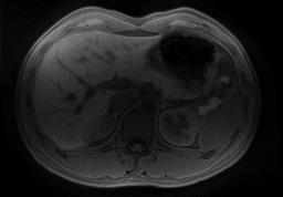 We are developing intelligent Bayesian MRI data acquisition using prior information
to vastly improve temporal and spatial resolution. This Bayesian MRI approach has
great potential for imaging moving organs such as the liver
(R21CA152275), enabling determination of liver cancer biomarkers including
transport parameters (R21DK090690).
We are developing intelligent Bayesian MRI data acquisition using prior information
to vastly improve temporal and spatial resolution. This Bayesian MRI approach has
great potential for imaging moving organs such as the liver
(R21CA152275), enabling determination of liver cancer biomarkers including
transport parameters (R21DK090690).
To see selected temporal and spatial 4D imaging publications
from our lab please visit here.