
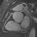
 The broad objective of this research is to develop magnetic resonance imaging (MRJ) techniques that will accurately detect significant lesions in the coronary arteries. MRI offers many advantages over CT and x- ray angiography for imaging coronary artery disease, including rich and adjustable contrasts between different tissues, noninvasive procedures, no use of ionizing radiation, and no risk of nephrotoxicity. Prior studies have demonstrated the feasibility of imaging both the coronary lumen and wall, but diagnostic accuracy is still impaired by motion artifacts. Coronary MR Angiography (MRA) technology is rapidly evolving. Studies in the prior funding cycle have led to several key findings. SSFP has been identified as the best sequence for coronary MRA because of its high SNR efficiency and fast imaging speed. Real time navigator gating provides higher spatial resolution and image quality than breath-holding for coronary MRA. There is still inconsistency in coronary MRA quality because of the use of electrocardiogram and the diaphragm navigator echo, neither of which directly measures coronary motion. We have recently developed the coronary fat navigator method that allows direct and accurate monitoring of coronary motion. Accordingly, we propose here to develop the coronary fat navigator for SSFP coronary MRA to achieve effective motion suppression. We have also developed a simultaneous multiple volume (SMV) algorithm for efficient navigator gating. Preliminary data demonstrate that the SMV algorithm shortens navigator gated scan time by 2-3 fold. Accordingly, we propose to incorporate SMV gating into navigator coronary MRA to shorten scan time. We have also obtained preliminary experience in imaging coronary vessel walls. The navigator technology can be directly employed to reduce motion blurring artifacts in high resolution plaque imaging, which would provide valuable information for characterizing atherosclerotic plaque in coronary arteries. This research consists of two specific aims: 1) develop an efficient and effective navigator method for imaging the coronary lumen and wall; and 2) evaluate the developed coronary MRI in patients with coronary artery disease by comparing to conventional x-ray angiography and intravascular ultrasound.