
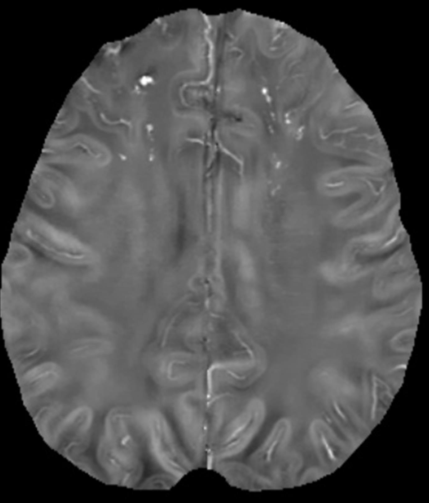
 The objective of this research is to develop a quantitative MRI method for mapping iron deposits in cerebral microbleeds (CMB). There is significant scientific and clinical interest in studying CMB, which are focal lesions of hemosiderin deposits from red blood cells leaked out of small brain vessels. CMB have become an important concern for managing stroke patients, particularly for assessing the risk of devastating intracerebral hemorrhage (ICH) in patients under anticoagulation. Quantitative mapping of iron deposits in CMB can be very valuable for stratifying risk of anticoagulation therapy. Currently, dark regions in T2* weighted MRI have been used to identify the presence of iron by interpreting the observed signal loss caused by the intravoxel dephasing effect of local magnetic fields of iron deposits. This hypointensity depends on voxel size and orientation, may be confused with other signal voids, and does not allow accurate quantification of iron. We propose a novel quantitative susceptibility mapping (QSM) approach to generate quantitative mapping of iron deposits using MRI by making full use of both phase and magnitude information in the T2* gradient echo image data. The phase image, typically neglected in MRI, is used to generate a local magnetic field map for fitting with susceptibility via the Maxwell equation. The magnitude image is used to extract tissue structure information for matching with susceptibility interfaces via least discordance. This morphology enabled dipole inversion approach is feasible for quantitatively mapping iron compound concentrations, as demonstrated in our preliminary studies, therefore enabling standardized and quantitative evaluation of CBM. Our proposed research consists of the following specific aims for developing and applying brain iron mapping technology. 1) Optimize data acquisition for mapping fields of CMB in the whole brain. 2) Develop a robust reconstruction for QSM of CMB iron deposits. 3) Validate QSM of bleeds and microbleeds in the brain using histological correlation. 4) Apply QSM to measure CMB in warfarin-treated patients with and without ICH.