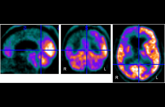
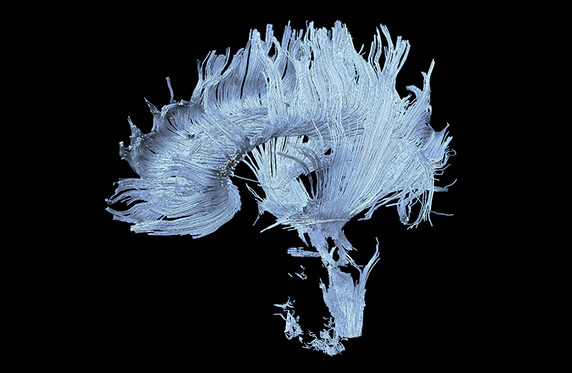
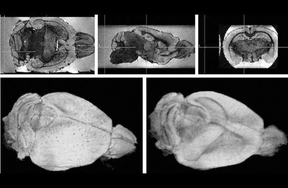
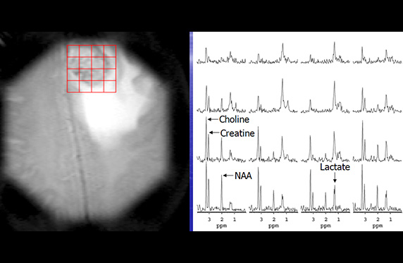
;
Center Description
The Citigroup Biomedical Imaging Center (CBIC) at Weill Cornell
Medicine is a state-of-the-art 15,000 square foot research facility housing the
Biomedical Imaging Core Facility of the College.
Major equipment includes two 3.0 Tesla magnetic
resonance imaging and spectroscopy (MRI/MRS) system; a 7.0 Tesla/30cm
bore pre-Clinical MRI/MRS system; a combined
positron emission tomography and computed tomography (PET/CT) system; a
pre-clinical MicroPET/SPECT/CT system; a multispectral optical imaging system,
a pre-clinical ultrasound system, a
medical cyclotron facility for production of radiotracers; and radiochemistry
laboratories for ligand synthesis.
The center has been carefully designed to facilitate complementary
use of all imaging modalities.
———————
Contact: Center Director, Douglas Ballon, Ph.D. (212) 746-5679


